
In contrast, the thick, porous peptidoglycan layer in the cell wall of Gram-positive bacteria gives greater access to antibiotics, allowing them to more easily penetrate the cell and/or interact with the peptidoglycan itself.

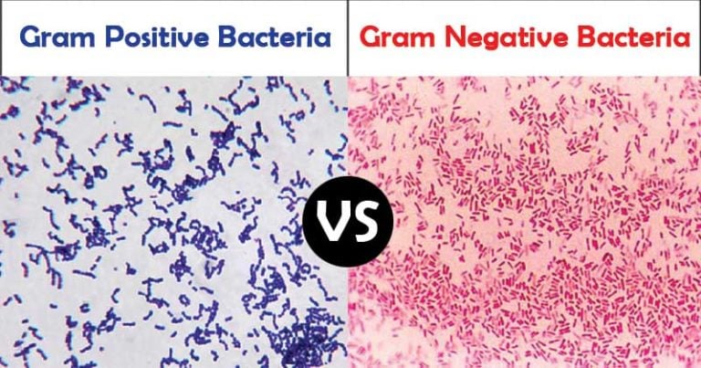
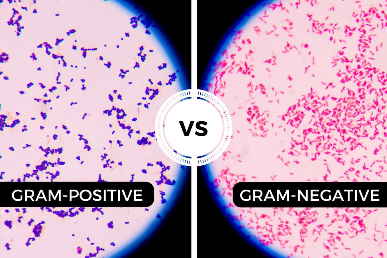
This second, outer membrane of Gram-negative bacteria is an effective barrier, regulating the passage of large molecules such as antibiotics into the cell. Figure 10 Arrangement of the cell wall in (a) Gram-positive and (b) Gram-negative bacteria. The inner membrane in (a) and (b) (shown as a double green line) is separated from the peptidoglycan layer by the periplasmic space. The epidemiology, microbiology, clinical manifestations, and treatment of gram-negative bacillary bacteremia will be reviewed here. What does it mean if bacteria are Gram positive The substance that retains the purple. 123 Bacteremia, in the strictest sense, refers to viable bacteria in the blood. Gram-positive bacteria have thick cell walls containing high levels of peptidoglycan, while gram-negative bacteria are characterized by thinner cell walls. Gram-negative bacillary sepsis with shock has a mortality rate of 12 to 38 percent mortality varies depending, in part, on whether the patient receives timely and appropriate antibiotic therapy 2-4. In the Gram-positive bacteria in (a) the peptidoglycan is a thick external layer shown in brown, while in the Gram-negative bacteria in (b) the peptidoglycan layer is much thinner and is surrounded by an outer membrane of lipopolysaccharide and protein (as a green wavy line). If the cells take on the red colour, they are Gram negative. They also cause different types of infections, and different. This diagram shows the differences in cell wall structure between Gram-positive and Gram-negative bacteria. Gram-positive and gram-negative bacteria stain differently because their cell walls are different.


 0 kommentar(er)
